

ICEECE2012 Poster Presentations Endocrine tumours and neoplasia (112 abstracts)
Can proteomic approach help us in diagnosis of Riedel’s thyroiditis? A case report
G. Donatini , P. Iacconi , L. Giusti , Y. Da Valle , F. Ciregia , G. Giannaccini , L. Torregrossa , A. Proietti , S. Mazzeo , F. Basolo & A. Lucacchini
University of Pisa, Pisa, Italy.
Background: Riedel’s thyroiditis (RT) is a rare thyroid disease. Clinical and citological differential diagnosis with thyroid malignancy is difficult pre-operatively and often only pathological report may confirm the diagnosis.
Methods: We report a case of a 72-year-old Italian woman with a known history of goiter, which showed a rapid increase in size at ultrasound check, suggesting malignancy. Based on non-diagnostic cytology (Thy 1), a total thyroidectomy was performed. Fine needle aspiration (FNA) of the removed thyroid was processed by two dimensional electrophoresis (2-DE) and the proteome compared both with anaplastic cancer and control samples (normal thyroid tissue). Protein spots found to be significantly diffenrently expressed were identified by western blot (WB) analysis using specific antibodies.
Results: The protein pattern of Riedel’s FNA revealed a superimposition with that of the control samples (Fig. 1). The comparison of RT FNA protein pattern with anaplastic thyroid cancer evidenced differentially expression of ferritin heavy chains, ferritin light chains and haptoglobins, as previously reported overexpressed in thyroid cancers. Therefore, we performed WB analysis of these proteins and we found that their expression were low or absent in RT and control samples despite the high concentrations present in FNA anaplastic samples.
Conclusions: The concurrent absence or low expression levels of haptoglobin, FLC and FHC in RT FNA sample strongly indicates the benign nature of the thyroid lesion. These results suggest the potential applicability of FNA proteome analysis for RT diagnosis and more generally to differentiate thyroid malignancy into thyroid nodule with non-diagnostic citology.
Declaration of interest: The authors declare that there is no conflict of interest that could be perceived as prejudicing the impartiality of the research project.
Funding: This research did not receive any specific grant from any funding agency in the public, commercial or not-for-profit sector.
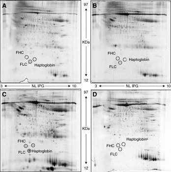
Figure 1 Proteomic analysis. 2-DE images. (A) Ridel’s thyroiditis (RT). (B) Control tissue (normal thyroid tissue). (C) Anaplastic thyroid cancer. (D) Control tissue.
}
Article tools
My recent searches
My recently viewed abstracts
Authors

Endocrine Abstracts
ISSN 1470-3947 (print) | ISSN 1479-6848 (online)
© Bioscientifica 2026 |
Privacy policy |
Cookie settings
BiosciAbstracts

Bioscientifica Abstracts is the gateway to a series of products that provide a permanent, citable record of abstracts for biomedical and life science conferences.
Find out more