

ECEESPE2025 ePoster Presentations Thyroid (198 abstracts)
Non-graves orbitopathy in a patient with hypothyroidism, hashimoto’s thyroiditis, and hiv on combined antiretroviral therapy
Naia Lataria 1 & Nino Rukhadze 2
1Todua Clinic, Tbilisi, Georgia; 2Tengiz Tsertsvadze Infectious Disease, AIDS and Clinical Immunology Center, Tbilisi, Georgia
JOINT635
Introduction: Hashimoto’s thyroiditis, also called chronic autoimmune thyroiditis, is one of the most common autoimmune thyroid diseases. The clinical manifestations can include either hypothyroidism or, in rare cases, hyperthyroidism. Thyroid Eye Disease (TED) occurs when thyroid autoantibodies cause inflammation in the connective tissue of the extraocular muscles and orbital fat, leading to clinical symptoms such as periorbital edema, lid retraction, and proptosis, among others. There is a close relationship between TED and thyroid-stimulating hormone receptor antibodies (TRAb). TED can also occur in patients with chronic autoimmune thyroiditis. HIV infects CD4+ T cells, and approximately 10–40% of patients receiving antiretroviral therapy (ART) may develop immune reconstitution inflammatory syndrome (IRIS).
Case Report: A patient was referred to an endocrinologist by an infectious disease specialist. HIV was diagnosed eight years ago, and the patient has been on combination antiretroviral therapy (Tenofovir, Dolutegravir, and Lamivudine) since then. Approximately two years ago, the patient was diagnosed with autoimmune thyroiditis and primary hypothyroidism but refused consultation with an endocrinologist.
On November 11, 2024, laboratory tests showed:
• TSH: >100 mIU/ml
• Anti-TPO: >600 IU/ml
Levothyroxine 75 mg was prescribed.
On January 8, 2025, follow-up laboratory results showed:
• TSH: 38.81 mIU/ml (reference range: 0.27–4.2)
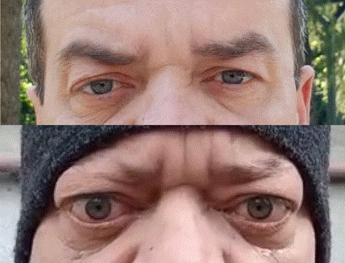
• FT4: 15.36 pmol/l (reference range: 12–22) Although there was a positive trend in thyroid function, the patient returned for consultation with signs of thyroid orbitopathy, more pronounced in the right eye, as well as spot baldness on the head and brow area—symptoms that were not present during the previous consultation two months prior. Before the endocrinology follow-up, the patient was evaluated by an ophthalmologist and was prescribed steroid eye drops. However, the patient did not notice any improvement in inflammation. On January 10, 2025, an MRI scan of the brain and orbits was performed, revealing:
• No acute focal organic pathology of the brain parenchyma
• Swelling of intraorbital fatty tissue in both eyes
• Exophthalmos in both eyes Levothyroxine dosage was increased to 100 mg, and Selenium 200 mg once daily was introduced for six months. Since Teprotumumab is not available in Georgia, intravenous methylprednisolone pulse therapy is being considered for further treatment.
Conclusion: This case highlights the importance of early ophthalmological evaluation in patients with autoimmune thyroiditis, including thyroid hormone assessment and thyroid autoantibody screening. It also underscores the need for early detection of eye complications, even in patients with autoimmune thyroiditis in the hypothyroid stage and without Graves’ disease.
}
Article tools
My recent searches
My recently viewed abstracts

Endocrine Abstracts
ISSN 1470-3947 (print) | ISSN 1479-6848 (online)
© Bioscientifica 2026 |
Privacy policy |
Cookie settings
BiosciAbstracts

Bioscientifica Abstracts is the gateway to a series of products that provide a permanent, citable record of abstracts for biomedical and life science conferences.
Find out more