

SFEBES2014 Oral Communications Clinical (6 abstracts)
11C-methionine PET--CT co-registered with volume MRI: a novel adjunctive imaging modality to aid diagnosis and management in patients with pituitary adenomas
Olympia Koulouri 1,2 , Narayanan Kandasamy 1 , Andrew Powlson 1,2 , Carla Moran 1,2 , Heok Cheow 2 , Nagui Antoun 2 , Miles Levy 3 , Andrew Hoole 2 , Krishna Chatterjee 1,2 , Neil Donnelly 2 , Richard Mannion 2 , Neil Burnet 1,2 , John Pickard 1,2 & Mark Gurnell 1,2
1University of Cambridge, Cambridge, UK; 2Addenbrooke’s Hospital, Cambridge, UK; 3University Hospitals of Leicester, Leicester, UK.
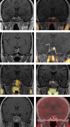
Although MRI remains the investigation of choice for pituitary imaging, it does not provide information about ‘functionality’ of lesions (e.g. residual adenoma vs post-surgical scar tissue) and cannot reliably identify all microadenomas.
We hypothesised that i) imaging with the PET ligand 11C-methionine, which is taken up at sites of peptide/protein synthesis, would permit more reliable identification of functioning pituitary adenoma and ii) co-registration of PET–CT with volume MRI (MetPETCT–MRI) would yield more accurate anatomical localisation of 11C-methionine uptake.
80 scans were performed in our centre between 2010 and 2013. MetPETCT–MRI was found to provide additional useful information in the following scenarios:
(A) Distinguishing residual functioning tumour from inactive or scar tissue post pituitary surgery (Figs 1 and 2) or medical therapy (e.g. cabergoline in prolactinoma) (n=29)
(B) Demonstrating medical therapeutic effects through significant reduction of tracer uptake post- compared to pre-treatment (Fig. 3) (n=15).
}
Article tools
My recent searches
My recently viewed abstracts
Authors

Endocrine Abstracts
ISSN 1470-3947 (print) | ISSN 1479-6848 (online)
© Bioscientifica 2026 |
Privacy policy |
Cookie settings
BiosciAbstracts

Bioscientifica Abstracts is the gateway to a series of products that provide a permanent, citable record of abstracts for biomedical and life science conferences.
Find out more