

Theranostics2016 4th Theranostics World Congress 2016 Spotlight on Neuroendocrine tumours (17 abstracts)
68Ga-Pentixafor-PET/CT for Imaging of Chemokine Receptor 4 Expression in Neuroendocrine Tumors – a head-to-head comparison with DOTATOC and FDG PET/CT
Michael Lassmann 1 , Rudolf A. Werner 1, , Alexander Weich 3 , Hans-Juergen Wester 4, , Michael Scheurlen 3 , Takahiro Higuchi 1, , Samuel Samnick 1 , Christina Bluemel 1 , Martina Rudelius 6 , Andreas K. Buck 1 , Constantin Lapa 1 & Ken Herrmann 1,
1Department of Nuclear Medicine, University Hospital Wuerzburg, Wuerzburg, Germany; 2Comprehensive Heart Failure Center, University Hospital Wuerzburg, Wuerzburg, Germany; 3Department of Internal Medicine II, Gastroenterology, University Hospital Wuerzburg, Wuerzburg, Germany; 4Scintomics GmbH, Fürstenfeldbruck, Germany; 5Pharmaceutical Radiochemistry, Technische Universität Muenchen, Munich, Germany; 6Institute for Pathology, University Hospital Wuerzburg, Wuerzburg, Germany; 7Department of Molecular and Medical Pharmacology, David Geffen School of Medicine at UCLA, Los Angeles, USA.
Introduction: Diagnostic imaging of neuroendocrine tumors (NETs) is the domain of somatostatin receptor (SSTR) agonists as well as FDG PET/CT in dedifferentiated tumors. SSTR also serves as target for receptor directed peptide therapy. More recently, specific ligands targeting the chemokine receptor 4 (CXCR4) were introduced potentially offering an additional theranostic option in NETs. Here we evaluated the CXCR4 expression using 68Ga-Pentixafor PET/CT in NET patients in comparison to 68Ga-DOTATOC and 18F-FDG PET/CT.
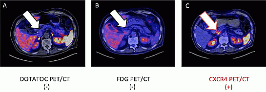
Material and methods: Eleven consecutive patients with histologically proven advanced NETs were retrospectively analyzed (three female; mean age, 69±10 years; Ki67 36±36%). 5/11 (45%) suffered from pancreatic NETs, 3/11 (27%) from ileum NETs, 2/11 (18%) from cancer of unknown primary and 1/11 (9%) was classified as a gastric NET. DOTATOC, FDG and Pentixafor PET/CT were performed in all patients within 4 weeks to confirm target expression of SSTR, CXCR4 and to detect dedifferentiated tumor lesions. Image analysis was performed visually. Immunohistochemical CXCR4 expression was evaluated in biopsy samples using monoclonal anti-human anti-CXCR4 antibodies.
Results: 7/11 (63%) initially presented with lymph node metastases, 3/11 (27%) with bone metastases, 9/11 (82%) with liver metastases, 2/11 (18%) with lung metastases and 1/11 (9%) with a brain metastasis. On visual analysis, Pentixafor was positive in 4/11 (36%), FDG in 9/11 (82%) and DOTATOC in 9/11 (82%) patients, respectively. Of the nine SSTR positive patients seven and three were also FDG- and CXCR4-positive. Two DOTATOC negative patients were FDG positive and one of them also Pentixafor positive. Three patients were positive on all three PET/CT scans. In 2/4 Pentixafor-positive patients, biopsy samples revealed intense CXCR4 expression.
Conclusions: In this pilot study, one third of NET patients were CXCR4 positive. However, one NET patient without SSTR expression was Pentixafor positive. Hence, CXCR4-directed radionuclide therapy can be envisioned for selected patients with SSTR-negative tumors.
}
Article tools
My recent searches
My recently viewed abstracts
Authors

Endocrine Abstracts
ISSN 1470-3947 (print) | ISSN 1479-6848 (online)
© Bioscientifica 2026 |
Privacy policy |
Cookie settings
BiosciAbstracts

Bioscientifica Abstracts is the gateway to a series of products that provide a permanent, citable record of abstracts for biomedical and life science conferences.
Find out more