BES2018 BES 2018 Pituitary metastasis from follicular thyroid carcinoma – a case report (1 abstracts)
Pituitary metastasis from follicular thyroid carcinoma – a case report
L Orioli 1 , E Fomekong 2 , T Duprez 3 , C Daumerie 1 & D Maiter 1
1Department of Endocrinology and Nutrition, Cliniques Universitaires Saint-Luc, UCL, Belgium; 2Department of Neurosurgery, Cliniques Universitaires Saint-Luc, UCL, Belgium; 3Department of Radiology and Medical Imaging, Cliniques Universitaires Saint-Luc, UCL, Belgium.
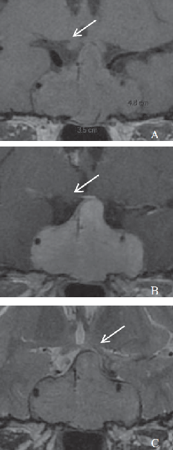
A 64-year-old Algerian woman was transferred to our institution with a pituitary tumor causing severe visual impairment. Magnetic resonance imaging (MRI) showed a T1-isointense, T2-hyperintense sellar mass measuring 48×35×30 mm, with homogenous enhancement after gadolinium injection (Figure 1). This mass extended to the suprasellar region, compressing the optic chiasma, and to both cavernous sinus, completely surrounding and narrowing carotid arteries. Ophthalmological examination showed near complete ophtalmoplegia due to bilateral palsies of VI and III nerves, bitemporal hemianopsia and bilateral optic nerve atrophy. Biological testing demonstrated complete anterior hypopituitarism with low prolactinemia and normal natremia (Table1). Medical history revealed that (i) a thyroid nodule corresponding to a follicular adenoma with signs of necrosis and hemorrhage had been resected in 2008, and (ii) that a non-functional pituitary tumor had been already diagnosed in Algeria two years before admission and incompletely resected by a trans-sphenoidal approach. The patient had developed postoperative corticotrope and thyrotrope insufficiency requiring treatment with hydrocortisone 20mg/day and levothyroxine 125 μg/day. Histopathological examination was compatible with a metastasis of a follicular variant of papillary thyroid carcinoma. A total thyroidectomy with central and laterocervical lymph node dissection was performed thereafter, confirming the presence of a 5 mm papillary microcarcinoma (pT1N0Mx). While thoraco-abdominal CT was negative, bone metastases of the left scapula and chondro-costal joints were demonstrated by a bone scintigraphy. Before planning a new pituitary surgery, a PET-CT was performed, showing a pituitary tumor without metabolic activity (SUV max 4.5) and without bone metastases. A transsphenoidal resection was then performed for biopsy and visual decompression but the intervention was complicated by abundant bleeding. The patient developed a transient diabetes insipidus post-operatively treated with desmopressin. Histopathological examination of the resected material revealed a metastasis of a follicular thyroid carcinoma. Tumoral cells were positive for TTF1, PAX8, thyroglobulin and TPO but negative for LH, FSH, GH, ACTH and TSH. The Ki67 proliferation index was estimated between 10% and 15%. Radioiodine ablation after recombinant human TSH administration was planned upon return of the patient in Algeria. Pituitary metastases from thyroid cancer are very rare. Less than 30 cases have been reported so far in the literature. In 11 cases, the thyroid cancer was a follicular thyroid carcinoma1. Most often, the metastases are part of a disseminated disease but in 9 cases it was the first sign of thyroid cancer1, as observed in our patient. As pituitary metastases from thyroid cancer tend to become large lesions with mass effects, symptoms most commonly include oculomotor palsies and visual deficit2. On the other hand, pituitary hormone deficiencies as well as hyperprolactinemia and diabetes insipidus are less frequently reported1 and seem to be more often associated with papillary and medullary rather than follicular thyroid carcinoma2. At MRI, pituitary metastases usually appear as iso- or hypo-intense lesions on T1-weighted images and hyper-intense on T2weighted image with marked enhancement after gadolinium injection1. Yet, these signs are not sufficient for diagnosis. As a consequence, neurosurgical biopsy or resection allows a definitive diagnosis as well as a primary treatment option especially when visual decompression is required. After neurosurgery and total thyroidectomy, if not performed earlier in the course of the disease, options include radioiodine (with or without recombinant human TSH) and electron beam radiotherapy1,3.

Figure 1 Magnetic resonance of the brain showing a large sellar and suprasellar tumor measuring 48×35×30 mm, iso-intense on T1-weighted images (A), enhancing after gadolinium injection (B), and moderately hyper-intense on T2-weighted images (C). The arrow shows the optic chiasma which is compressed on the left side. Both cavernous sinuses are largely invaded.
| Biological testing | Normal values | Results |
| ACTH | 5.0–49.0 pg/ml | 3.6 |
| Cortisol | 130.0–500.0 nmol/l | 1.5 |
| TSH | 0.27–4.2 mU/l | <0.01 |
| Free T4 | 12.0–22.0 pmol/l | 28.8 |
| GH | <4.0 ng/ml | <0.1 |
| IGF-1 | 100.0–232.0 ng/ml | 42.6 |
| LH | 7.7-15.5 UI/l$ | 0.3 |
| FSH | 25.8–134.8 UI/l$ | 0.9 |
| Estradiol | <5.0–54.7 ng/l$ | <12 |
| Prolactin | 5.0–23.0 μg/l | 3.2 |
| Natremia | 135–145 mmol/l | 143 |
References: 1. Barbaro D, Desogus N, Boni G. Pituitary metastasis of thyroid cancer. Endocrine. 2013 Jun;43(3):485–93.
2. Vianello F, Mazzarotto R, Taccaliti A et al. Follicular thyroid carcinoma with metastases to the pituitary causing pituitary insufficiency. Endocrinol Diabetes Metab Case Rep. 2013;2013:130024.
3. Prodam F, Pagano L, Belcastro S et al. Pituitary metastases from follicular thyroid carcinoma. Thyroid. 2010 Jul;20(7):823–30.
 }
}


