EU2019 Society for Endocrinology: Endocrine Update 2019 Poster Presentations (73 abstracts)
Pituitary abscess with meningitis: a rare presentation
Rupinder Kochhar , Asma Naseem & Tara Kearney
Department of Endocrinology, Salford Royal Hospital, Salford, UK.
Case history: A 42-year-old lady was referred from neurology clinic after being assessed for symptoms of headaches, dizziness, transient visual problems and paraesthesia over her limbs for 2 months. On review, she complained of amenorrhoea and was noted to be pale, however, her neurological examination including visual fields to confrontation and ocular movements were normal. Subsequent investigations were consistent with pan-hypopituitarism and she was promptly commenced on hydrocortisone.
Investigations:: LH: 1.0 U/L (2.0–13.0)
FSH: 4.7 U/L (3.0–10.0)
Estradiol: <44.0 pmol/L
TSH: 0.04 mU/L (0.35–5.5)
Free T4: 10.0 pmol/L (10–20)
Prolactin: 684 mU/L (59–619)
Cortisol: 20 nmol/L (200–500)
Sodium: 124 mmol/L (133-146)
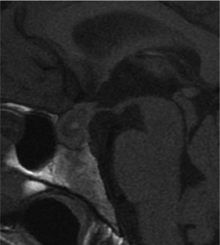
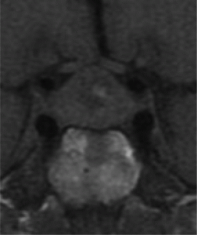
She had an urgent MRI scan which revealed 18 × 14 mm pituitary mass with minor optic-chiasmal compression and unusual signal characteristics (Image 1.0/1.1).

Image 1.0

Image 1.1
Results and treatment: Two weeks later she presented to emergency with worsening headache, persistent vomiting and visual blurring. She was noted to have slightly reduced visual acuity in the left eye, however, visual fields were normal. Urgent CT scan did not reveal acute haemorrhage and she was being considered for elective surgery and a high-resolution MRI Pituitary was planned. Whilst admitted she developed a severe headache, photophobia and vomiting. Lumbar puncture revealed raised intracranial pressure & lymphocytic pleocytosis.
CSF results:: Opening pressure: 43 cm H2O
Protein: 1.2 g/L (0.08–0.32)
WCC: 2000 (P 35%, L 65%)
Culture/Virology/AFB/Mycobacterium spp. PCR: Negative
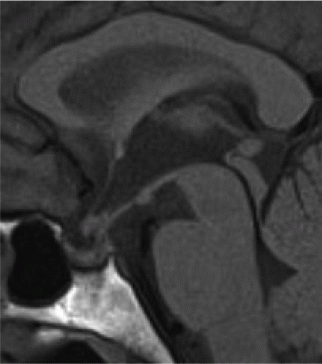
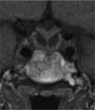
She was then started on i.v. Ceftriaxone & Acyclovir and review of her images at regional Pituitary MDT raised suspicion of infective pathology. She was being considered for urgent trans-sphenoidal debulking/decompression, however, she subsequently improved clinically and repeat imaging showed shrinkage of the pituitary lesion with infundibular thickening. She also developed cranial diabetes insipidus and was managed with oral desmopressin. She was managed conservatively for a pituitary abscess and completed an 8-week course of antibiotics. Repeat imaging at 2 and 7 months showed complete resolution of the lesion (Image 2.0/2.1) and she remains on pituitary hormone replacement.

Image 2.0

Image 2.1
Conclusions and points for discussion: A pituitary abscess is a rare cause for a sellar mass which may present without classical symptoms of infection. Atypical imaging characteristics should raise the index of suspicion.
What are the prospects of recovery of pituitary function?
What should be the follow-up surveillance?


