ICEECE2012 Poster Presentations Thyroid (non-cancer) (188 abstracts)
Decreased echogenicity and increased vascularization of fetal thyroid on 2D-ultrasonography caused by mother’s Graves’ disease
M. Gietka-Czernel 1 , M. Debska 1 , P. Kretowicz 1 , H. Jastrzebska 1 & G. Wasniewska 2
1Medical Center of Postgraduate Education, Warsaw, Poland; 2Bielanski Hospital, Warsaw, Poland.
Fetal ultrasonography is recommended in pregnant women with elevated TSH receptor antibodies (TRAK) or treated with antithyroid drugs in attempt to recognize fetal thyroid dysfunction. We report two cases of pregnant women with Graves’ disease in whom fetal thyroid ultrasonography was especially useful in diagnosing child involvement.
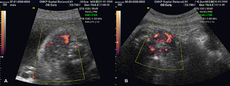
A 33-year-old woman at 30-week pregnancy with 3-months history of hyperthyroidism treated with propylothiouracil (PTU) 150 mg daily. Patient TSH was 0.033 μIU/ml (normal, 0.4–4.0 μIU/ml), fT4 9.24 pmol/l (normal, 11.5–22.7 pmol/l), fT3 6.1 pmol/l (normal, 2.8–6.5 pmol/l), TRAK 17.28 IU/ml (positive results >1.8 IU/ml). Fetal ultrasonography identified enlarged hypoechoic thyroid gland with increased peripheral vascularization. Fetal hormones concentrations obtained through cordocentesis were indicative for subclinical hypothyroidism: TSH 18.5 μIU/ml (normal, 2.4–12.8 μIU/ml), fT4 11.0 pmol/l (normal, 9.7–16.7 pmol/l), fT3 1.17 pmol/l (normal, 1.1–3.7 pmol/l).
The second patient, 30-year-old woman at 35-week pregnancy with 5 months history of hyperthyroidism and inadequate PTU treatment. Her TSH was <0.001 μIU/ml, fT4 27.8 pmol/l, fT3 18.1 pmol/l, TRAK 35.5 IU/ml. Fetal ultrasonography revealed hypoechoic goiter with increased central vascularization. The diagnosis of the mother and fetal hyperthyroidism was established.
Our results show that fetal thyroid gland when affected by transplacental passage of of maternal TRAK can demonstrate the same characteristic ultrasound pattern of Graves’ disease as in adults: enlargement, hypoechogenicity and hypervascularization. In accordance to previous observation the mode of blood flow within fetal thyroid enables discrimination between fetal hyperthyroidism and hypothyroidism caused by ADT overdosing. The 2D- ultrasound images of fetal goiter with increased blood flow are demonstrated.
Decreased echogenicity, increased thyroid size and increased peripheral vascularization of the fetal thyroid (A); decreased echogenicity, increased thyroid size and increased central vascularization of the fetal thyroid (B).
Declaration of interest: The authors declare that there is no conflict of interest that could be perceived as prejudicing the impartiality of the research project.
Funding: This research did not receive any specific grant from any funding agency in the public, commercial or not-for-profit sector.

 }
}


