

ICEECE2012 Poster Presentations Diabetes (248 abstracts)
Oxidative stress and histopathological differences on the skin of diabetes mellitus compromised rats
T Andrade 1 , V Malachias 2 , G Caetano 3 , D Masson 1 , A Jordão 1 , C Landim 1 & M Frade 1
1University of Sao Paulo, Ribeirao Preto, Brazil; 2Londrina State University, Londrina, Brazil; 3University of Sao Paulo, Sao Paulo, Brazil.
Background: Several studies on diabetes mellitus and cutaneous wounds have been reported. However, only few studies show the influence of diabetes on the skin regarding its oxidative stress, antioxidants mechanisms and histopathological aspects.
Objectives: The aim of this study was to evaluate these parameters by comparing them to non-diabetic rats skin.
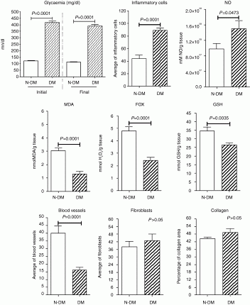
Methods: Ten Wistar rats were assigned to groups: diabetic rats (induced by streptozotocin 45 mg/kg; DM; n=5) and non-diabetic rats (N-DM; n=5). After verifying the diabetes status (hyperglycemia), animals were euthanized and biopsies were taken from the dorsal surface using a skin biopsy punch (1.5 mm diameter). Biochemical analysis was carried out to measure markers of oxidative stress (MDA, malondyaldeide; NO, nitric oxide; FOX, H2O2) and antioxidant activity (GSH, glutathione). Histological analysis (HE and Gomori’s trichrome staining) was performed to assess inflammatory cells, blood vessels, fibroblasts and the percentage of collagen area.
Results: Hyperglycemia was confirmed for DM group, reaching statistical difference compared to N-DM group (P=0.0001). Diabetes pathology increased the number of inflammatory cells (P=0.0001) and NO production (P=0.0473) on the skin, however, it decreased MDA (P=0.0001), FOX (P=0.0001) and GSH (P=0.0035). The number of blood vessels was lower for the DM group compared to the N-DM group (P=0.0001), suggesting that there is an influence of diabetes pathology on the quantity of skin blood vessels. The number of fibroblasts an collagen content were similar for both groups (P>0.05).
Conclusion: The pathophysiology of diabetes changed the skin by increasing the influx of inflammatory cells and NO production, decreasing the antioxidant defense and the number of blood cells.
Glycaemia (mg/dl) of diabetic (DM) and non-diabetic (N-DM) rats. Distribution of variables inflammatory cells, NO, MDA, FOX, GSH, blood vessels, fibroblast and collagen of dorsal skin in diabetic (DM) and non-diabetic (N-DM) rats.
Declaration of interest: The authors declare that there is no conflict of interest that could be perceived as prejudicing the impartiality of the research project.
Funding: This work was supported, however funding details unavailable.
}
Article tools
My recent searches
My recently viewed abstracts
Authors

Endocrine Abstracts
ISSN 1470-3947 (print) | ISSN 1479-6848 (online)
© Bioscientifica 2026 |
Privacy policy |
Cookie settings
BiosciAbstracts

Bioscientifica Abstracts is the gateway to a series of products that provide a permanent, citable record of abstracts for biomedical and life science conferences.
Find out more