ECEESPE2025 ePoster Presentations Pituitary, Neuroendocrinology and Puberty (220 abstracts)
A case of leydig cell tumor presenting with peripheral precocious puberty
Emine Şeyma Eken 1,2 , Meltem Korkmaz Vural 1,2 , Keziban Aslı Bala 1,3 , Firat Serttürk 1,4 , Erdal Kurnaz 1,2 , Melikşah Keskin 1,2 , Halit Üner 1,5 , Şule Yeşil 1,6 & Şenay Savaş Erdeve 1,2
1Ankara Etlik City Hospital, Pediatric Endocrinology Clinic, Ankara, Türkiye; 2Ankara Etlik City Hospital, Pediatric Endocrinology Clinic, Ankara, Türkiye; 3Ankara Etlik City Hospital, Ankara, Türkiye; 4Ankara Etlik City Hospital, Pediatric Surgery clinic, Ankara, Türkiye; 5Ankara Etlik City Hospital, Pathology Clinic, Ankara, Türkiye; 6Ankara Etlik City Hospital, Pediatric Oncology Clinic, Ankara, Türkiye.
JOINT2753
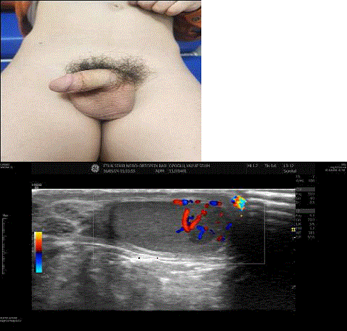
Introduction: Leydig cell tumors are sex cord-stromal gonadal tumors originating from Leydig cells. Although rare, accounting for 1-3% of all testicular tumors, they are the most common hormone-secreting testicular tumors. These tumors have been found to be associated with peripheral precocious puberty. In this study, we present a case of Leydig cell testicular tumor in a patient presenting with peripheral precocious puberty. Case:A 5-year-and-12-month-old male patient presented to our outpatient clinic with complaints of pubic hair growth. His medical history revealed that he had no known chronic illnesses, hospitalizations, or medication use. On physical examination, his height was 125.2 cm (2.64 SDS), body weight was 32.9 kg (3.22 SDS), body mass index was 20.99 kg/m2 (2.66 SDS), and target height was 175 cm (-0.19 SDS). His genetic alignment was +2.83 SDS. The right testis measured 6 mL, the left testis measured 4 mL, and his stretched penile length was 7 cm. Pubic hair was noted at Tanner stage 3, and axillary hair was also present (Image 1). The patient’s complete blood count, biochemistry, and thyroid function tests were normal. His FSH was <0.3 IU/l(0.2-2.1), LH was 0.36 IU/l(0.1-1.3), total testosterone was 301 ng/dl (2.5-32), and AFP and β-HCG were negative. His bone age was consistent with 11 years. Scrotal imaging showed the right testis measured 24×14×14 mm, the left testis measured 15×10×11 mm, and a lesion with increased vascularity was noted in the right testis parenchyma, measuring 14x7x12 mm, presenting as a hypoechoic area resembling a mass (Image 2). An LH-RH stimulation test revealed a peak LH of 0.85 IU/land peak FSH of 1.21 IU/l, which was consistent with peripheral precocious puberty. The patient underwent a frozen-section guided right testis preserving mass excision, and the pathological diagnosis was Leydig cell tumor consisting of large, eosinophilic, and granular cytoplasm cells, measuring 1.2 cm in diameter. The surgical margins were reported as intact. Follow-up scrotal and abdominal ultrasonography performed one month later were normal, and subsequent gonadotropin tests showed FSH: 4.72 IU/l(0.2-2.1), LH: 3.89 IU/l(0.1-1.3), and total testosterone: 108 ng/dl (2.5-32). Considering the possibility of central precocious puberty, GnRH analog treatment was started.

Conclusion: Leydig cell tumors are a cause of peripheral precocious puberty, and these patients typically present with a testicular mass, pubic hair growth, elevated testosterone, and low gonadotropin levels. Scrotal imaging is essential in these cases. With testis-preserving surgery, the prognosis is generally favorable, but patients should be carefully monitored for the potential development of central precocious puberty.
 }
}


